Cryo-Electron Microscopy
We use cryo-electron microscopy to answer questions about the form and function of biological "machines" that play key roles in human health and disease.

Advances in microscope technology and computing have paved the way for cryo-electron microscopy to move structural biology into a new era — allowing scientists to study the form and function of biological "machines" that are too large to study using X-ray crystallography.
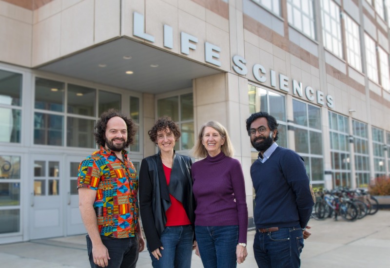
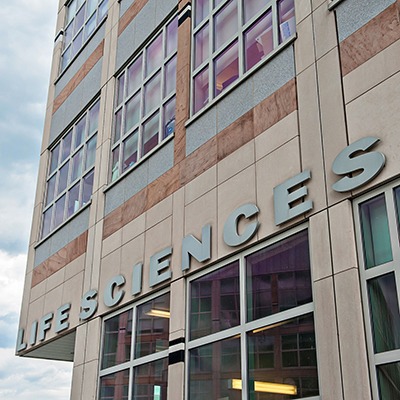
The U-M Life Sciences Institute is home to a world-class electron microscopy facility, with state-of-the-art instruments and faculty specializing in this emerging field.

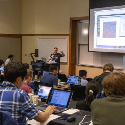
New funding through the U-M Biosciences Initiative will enable the university to become an international leader in the field and a premier destination for cryo-EM research and training. With this funding, the cryo-EM program is expanding into new technologies and developing additional educational and training opportunities to bring the technique to labs across campus, as well as to the growing ranks of practitioners across the world.
Registration has opened for the 2026 Cryo-Electron Tomography Workshop. Applications are due March 20, 2026. Learn more and apply.
We want to help researchers at U-M perform the best structural biology possible.
Use our access form to submit a brief description of the sample you would like to visualize. Based on your request, our faculty will schedule one-on-one meetings to discuss strategies.