


Newly identified protection mechanism serves as first responder to cellular stress

Researchers at the University of Michigan Life Sciences Institute have identified a new type of rapid-response defense mechanism that helps protect cells from environmental stress while giving slower, well-known protection systems time to act.
“It’s like a first responder rushing to an alarm while the larger response team mobilizes,” says Natsuko Jin, Ph.D., a postdoctoral research fellow in the lab of LSI faculty member Lois Weisman, Ph.D., and the lead author of a study published June 21 in the Journal of Cell Biology.
Generally, when cells are put under stress, adaptation mechanisms kick in. They trigger transcriptional machinery and, through gene expression, the cell produces new proteins to respond to the stress and keep itself alive.
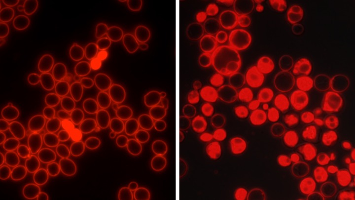
In yeast, a single-cell organism often used to study fundamental cellular biology, a much faster type of response was also observed — an immediate and short-lived spike in the production of a signaling lipid that is usually seen only in miniscule quantities.
When the scientists short-circuited the yeasts’ ability to generate this rapid response, the yeast succumbed to an environmental stress at catastrophic rates.
“This is the first time an early protection pathway that works faster than gene expression has been identified,” Jin says. “Since many of the key players have been preserved by evolution up into people and other mammals, our investigations suggest this and other types of early protection pathways may exist more broadly, and they may respond to different types of cellular stress.”
For this study, the yeast were put into an environment with a high concentration of salt — what scientists call high osmolarity. Within a few minutes, each cell responds by setting off a signaling cascade that activates a key protein kinase — Hog1 — which travels from the cell’s cytoplasm into the nucleus, where it promotes changes in gene expression. These changes in gene expression take between 30 minutes and an hour to start to have an effect, and up to two hours to be fully activated.
Meanwhile, the researchers also observed a sharp, immediate spike in a signaling lipid known as PI3,5P2, which is produced by an organelle called the vacuole in yeast. The yeast vacuole is similar to the lysosome in complex organisms.
“Within one minute you see a five-fold elevation of this lipid,” says Weisman, the senior author on the paper and a professor of cell and developmental biology at the U-M Medical School. “Within five minutes, it’s a 20-fold increase. Then, without us doing anything else to the cells, it plateaus and drops off.”
When regular yeast were put into this high salt, or hyperosmotic, environment for four hours, most did just fine.
When the researchers used genetic manipulation to knock out the well-known, longer-term response pathway that produces Hog1, 30 percent of the cells died.
“Still, 70 percent did just fine,” Weisman notes.
But when they removed the cell’s ability to produce PI3,5P2, 80 percent died.
“So we know it’s doing something protective before the gene expression kicks in,” she adds. “If they don’t have it, most die.”
Exactly how PI3,5P2 conveys a benefit to the cell is not yet understood, Jin notes. The current study examined the upstream regulators of the signaling lipid, and demonstrated they were distinct both in time and space from the action of the Hog1 pathway.
She also notes that while the observation that PI3,5P2 spikes under hyperosmotic conditions dates back to the late 1990s, its role had previously been unclear. Jin’s investigation started with the desire to understand what causes the spike and what physiological role it might play.
Go to Article
Early protection to stress mediated by CDK-dependent PI3,5P2 signaling from the vacuole/lysosome, Journal of Cell Biology. DOI: 10.1083/jcb.201611144