


Research highlight: Chemical tool illuminates pathways used by dopamine, opioids and other neuronal signals
University of Michigan researchers have developed a new tool to better understand how chemicals like dopamine and epinephrine interact with neurons.
These chemicals are among a wide variety of signals that get processed in the brain through G protein-coupled receptors (GPCRs), proteins that sit on the surface of neurons to receive messages — in the forms of proteins, sugars, fats, even light — that inform cellular behavior.
GPCRs are involved in an enormous number of biological functions, making them a prime target for treating diseases; more than one-third of FDA-approved drugs target GPCRs. But to fully understand how various molecules interact with GPCRs, researchers need to be able to detect those molecules across the whole brain with high spatial resolution.
“The challenge in our field has been achieving the right balance between a detailed view and the whole picture across the brain,” says Wenjing Wang, Ph.D., a neuroscientist at the U-M Life Sciences Institute.
“With most existing tools, you can detect a neural modulator either in a small part of the brain with high spatial resolution or in the whole brain with very low resolution,” adds LSI faculty member Peng Li, Ph.D. “But we need to identify the cells that respond to the neuromodulators across various brain regions, in high resolution.”
In a new study published April 22 in the Proceedings of the National Academy of Sciences, Wang, Li and their colleagues unveiled a new chemical tool that achieves both goals for three chemicals that all target GPCRs.
Ideally, we aim to be able to create a brain map for multiple neuromodulators concurrently, offering a comprehensive understanding of the sites of neuromodulation.
Wang’s lab at the LSI uses protein engineering to develop technologies that can detect how signaling molecules travel within the brain to reach and interact with specific neurons. They previously created a tool to reveal the presence of opioids, another GPCR binding partner, at a cellular level.
When the molecule is detected, the tool creates a permanent fluorescent mark in the cells. Thus, researchers can see the specific cells that are highlighted, as well as the whole picture of cells across the brain.
This latest work broadens the utility of that sensor to detect multiple types of GPCR activators, beyond just opioids. So far, the team has tested the tool with opioids and epinephrine in cultured neurons and in mouse models. The team also expanded the tool to use both green and red fluorescence, enabling the tracking of multiple molecules at once.
“Coming from detecting just opioids, we now have a tool that we can begin to easily modulate for various signals that interact with GPCRs,” says Wang, who is also an assistant professor of Chemistry in the U-M College of Literature, Science, and the Arts. “The goal is eventually to even study the interplays of different signaling pathways simultaneously.”
The team cautions that while the tool provides important visualizations of how signals travel across neurons for analysis postmortem, it cannot be used to track chemicals in real time, as it takes several hours for the fluorescence to appear. But it does offer a new path forward for improving understanding of neuronal signaling and the role of GPCRs as drug targets.
“Ideally, we aim to be able to create a brain map for multiple neuromodulators concurrently, offering a comprehensive understanding of the sites of neuromodulation,” says Li, who is also an assistant professor in the U-M School of Dentistry.
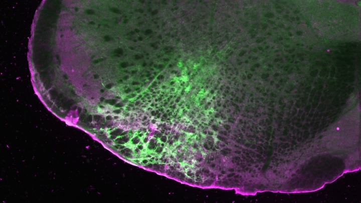
Top image: A slice of the mouse brainstem demonstrates the detection of morphine (green) and the expression of the SPOTIT sensor (magenta). Image credit: Noam Gannot and Peng Li, U-M Life Sciences Institute.
Go to Article
“Single-chain fluorescent integrators for mapping G-protein-coupled receptor agonists,” Proceedings of the National Academy of Sciences. DOI: 10.1073/pnas.2307090121